top of page
Element 1.1 rehabilitated with post-extraction Prama implant immediately loaded Neck
Dr. Roberto Luongo
A 26-year-old patient came to my observation for a traumatic fracture of element 1.1. A prosthetically guided implant surgery is planned, taking into account the final position of the dental element, in view of a contextual orthodontic treatment with removable aligners. The tooth was extracted in an atraumatic manner and a 4.2 x 13 mm Prama RF implant inserted. At the same time, the vestibular tissue was first thickened by means of a connective graft taken from the maxillary tuberosity, after having been deprived of the epithelial component, then a particulate bone graft of porcine origin was inserted between the implant and the vestibular bone crest. Finally, a screw-retained temporary non-functional immediate loaded crown was inserted.
After 6 months, an optical impression was taken (Carestream 3600) and a final screw-retained crown made of zirconia. From the CT cone beam 1 year after the extraction, bone regeneration was evident right up to the anodized neck of Prama implant.


Image courtesy: Dr. Roberto Luongo
Pre-op clinical image

Image courtesy: Dr. Roberto Luongo
Detail of pre-op clinical image
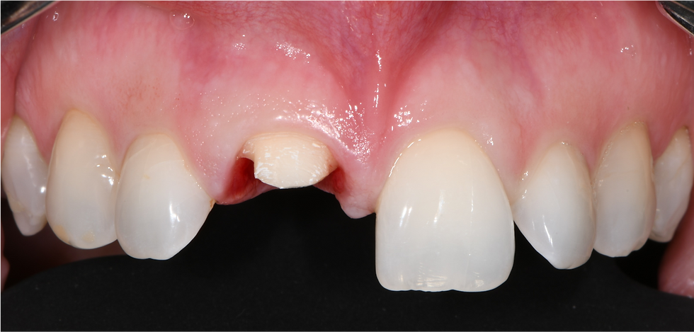
Image courtesy: Dr. Roberto Luongo
Traumatic fracture of element 1.1
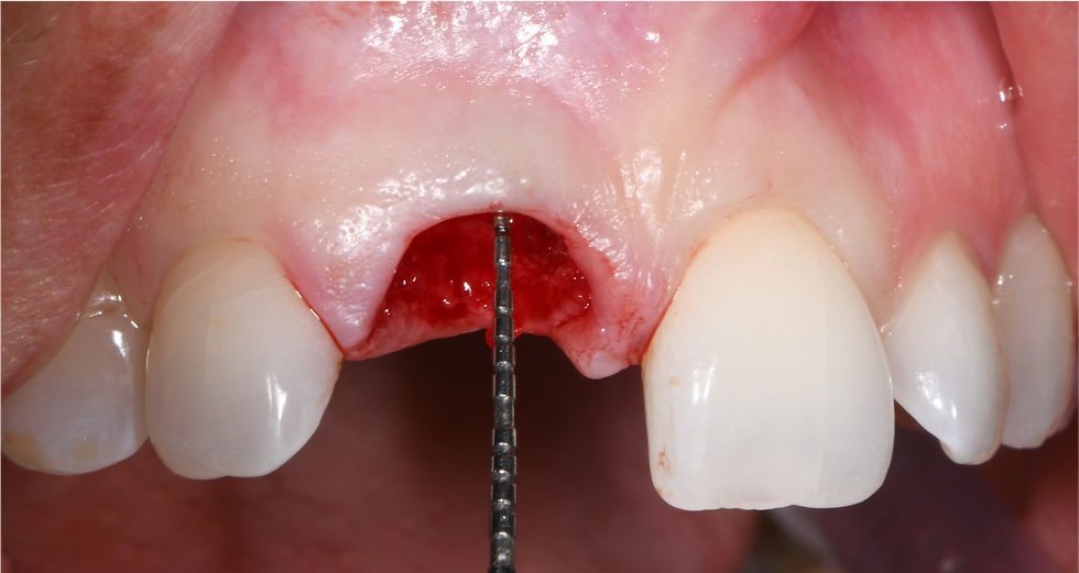
Image courtesy: Dr. Roberto Luongo
Extraction of the hopeless element

Image courtesy: Dr. Roberto Luongo
Planning of the prosthetic-guided surgery
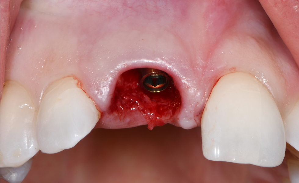
Image courtesy: Dr. Roberto Luongo
Implant placed in the post-extraction socket

Image courtesy: Dr. Roberto Luongo
At the same time, a connective graft is taken from the maxillary tuberosity
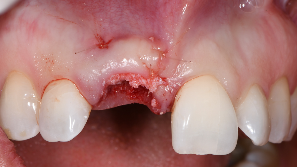
Image courtesy: Dr. Roberto Luongo
The gap is filled with a particulate bone graft of porcine origin

Image courtesy: Dr. Roberto Luongo
Immediate loading of the temporary crown

Image courtesy: Dr. Roberto Luongo
X-ray control at the moment of prosthetic loading
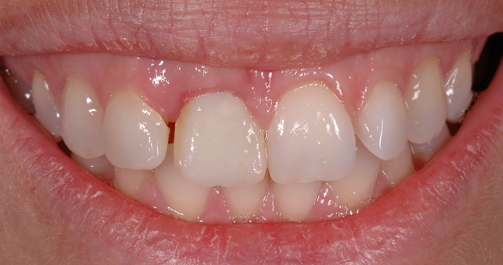
Image courtesy: Dr. Roberto Luongo
3 months clinical follow-up

Image courtesy: Dr. Roberto Luongo
6 months clinical follow-up
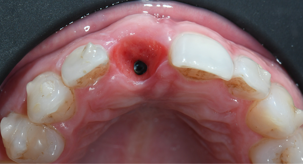
Image courtesy: Dr. Roberto Luongo
Occlusal detail of the healing after 6 months
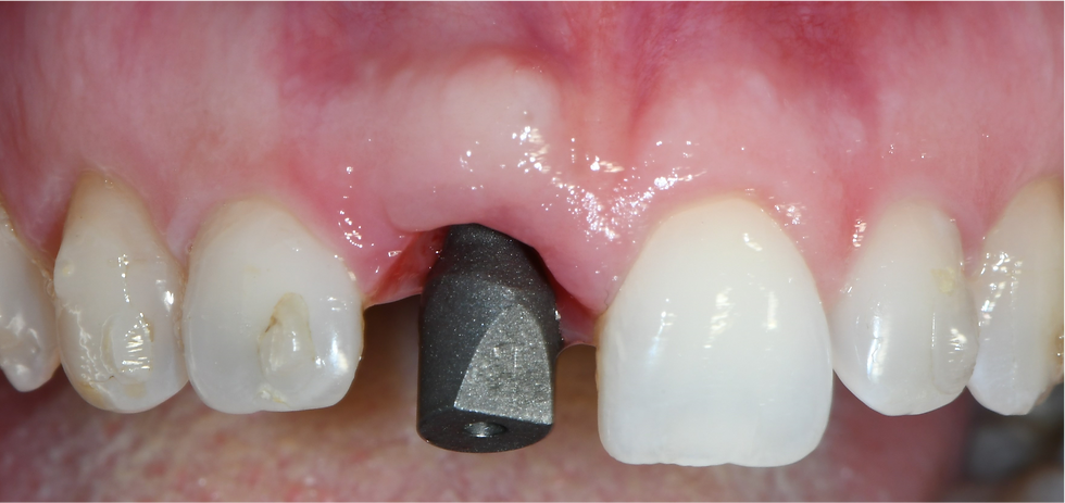
Image courtesy: Dr. Roberto Luongo
Impression taking with intraoral scanner
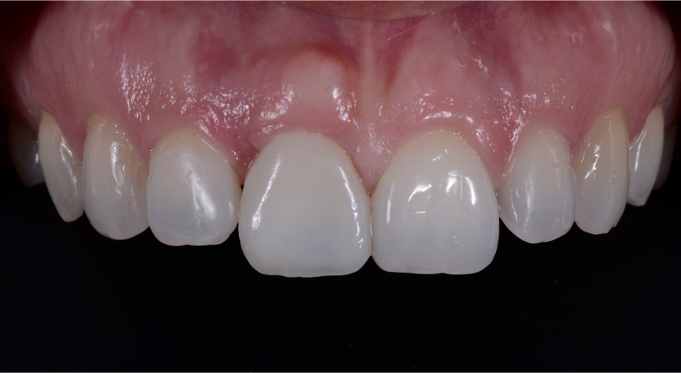
Image courtesy: Dr. Roberto Luongo
Final zirconia screw-retained crown
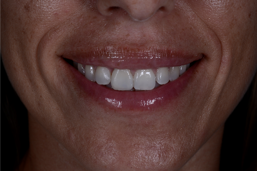
Image courtesy: Dr. Roberto Luongo
Final smile

Image courtesy: Dr. Roberto Luongo
X-ray one-year control
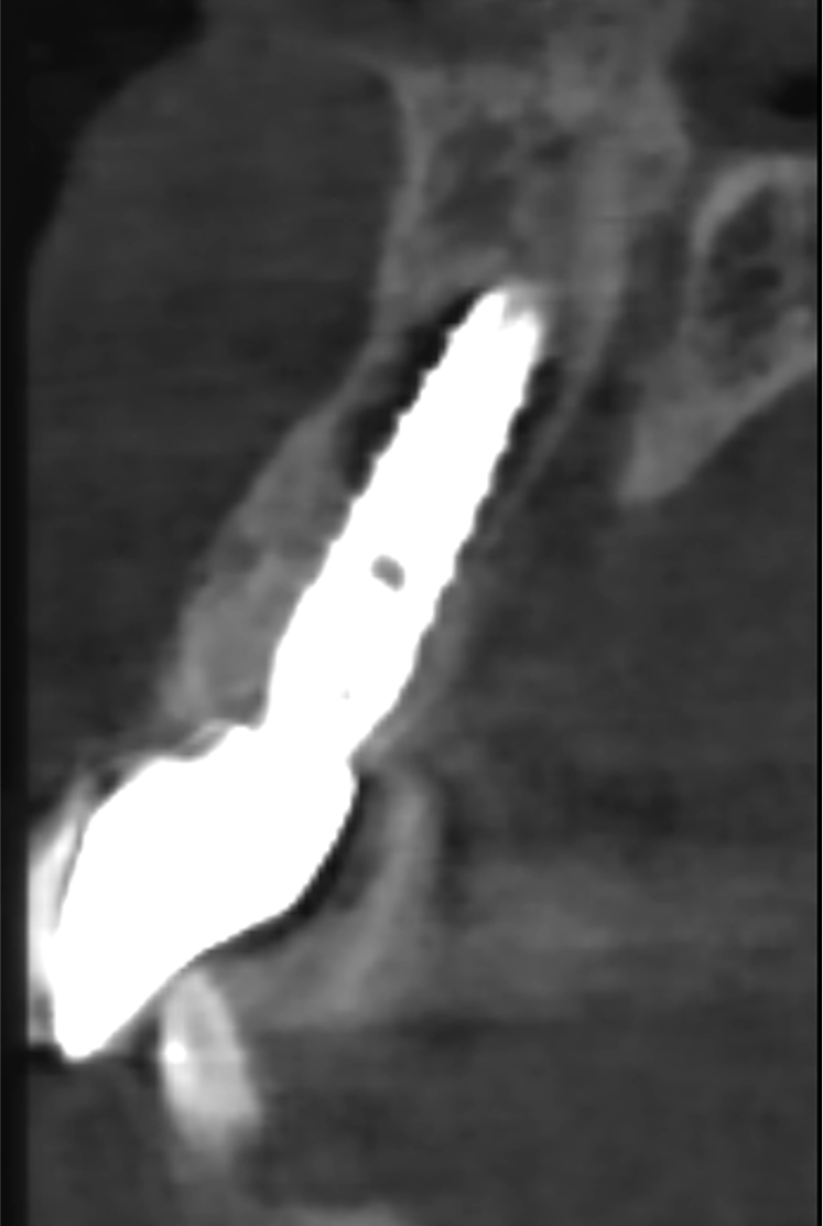
Image courtesy: Dr. Roberto Luongo
X-ray one-year control
bottom of page