Element 1.4 rehabilitation after ridge preservation technique
Prof. Antonio Barone, Geneva, Dr. Fortunato Alfonsi, Domodossola (VCO)
DT Marco Stoppaccioli, L’Aquila
The patient came to our observation showing a radicular fracture on the element 1.4. It was decided to proceed with the extraction of the element and a socket preservation, which allowed us to maintain the volumes of the alveolus.
“The decision to face this case with Prama was inspired by the need, in a site like this with a high aesthetic value, to maintain, optimize and stabilize the soft tissue structure. The Prama IN healing abutments, which embrace the implant neck, have contributed to shaping the tissue profile, to maintain the biological width, a concept from which we start in designing the emergence profile of the crown. Prama makes simple the clinician’s daily work.”
cit. Prof. Antonio Barone, Dr. Fortunato Alfonsi and DT Marco Stoppaccioli
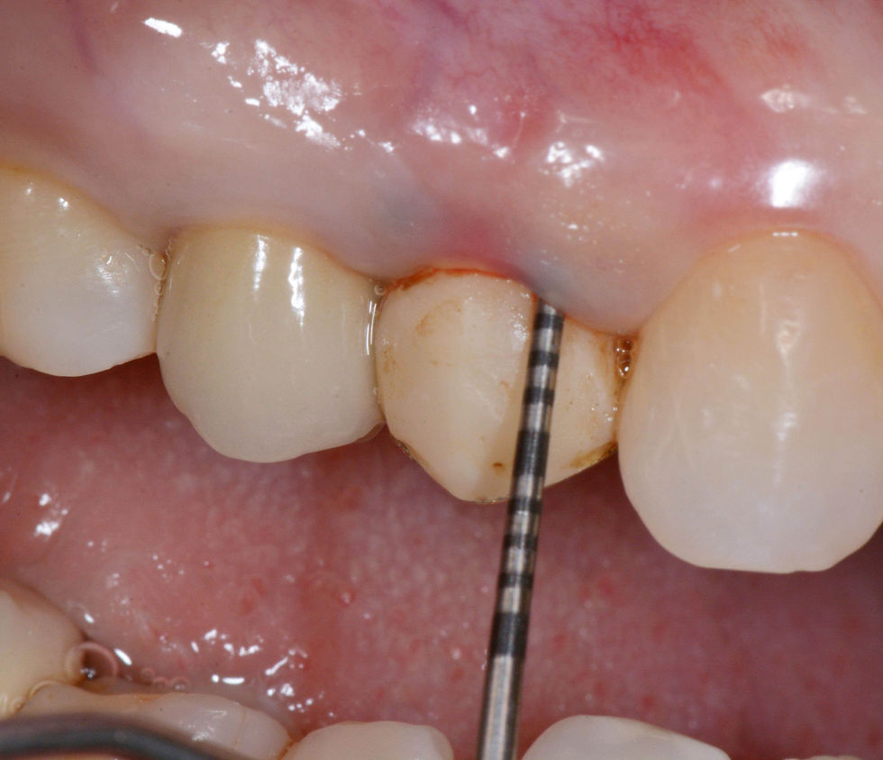
Initial situation.

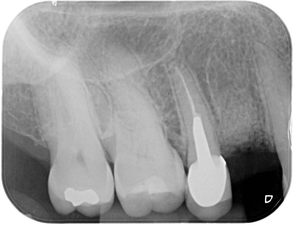
Pre-operative intraoral radiograph of the element 1.4.

Occlusal view of the post-extraction socket with evidence of loss of the vestibular bone wall.

A socket preservation technique is chosen to preserve the alveolar bone volumes, carried out with -tricalcium phosphate and a collagen membrane with a fibrin sponge covering the alveolus.

A socket preservation technique is chosen to preserve the alveolar bone volumes, carried out with ß-tricalcium phosphate and a collagen membrane with a fibrin sponge covering the alveolus.

A socket preservation technique is chosen to preserve the alveolar bone volumes, carried out with -tricalcium phosphate and a collagen membrane with a fibrin sponge covering the alveolus.

The alveolus is sutured to induce a healing by second intention.
Radiographic healing of the alveolus after 6 months.
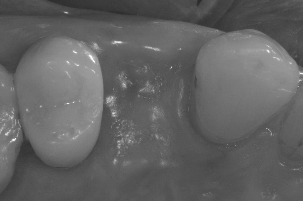
Clinical healing of the post-extraction site after 6 months.
Insertion of the Prama RF implant. Then a Prama IN healing abutment will be placed: it surrounds the implant neck by 0.5 mm.
Clinical healing after 6 months.

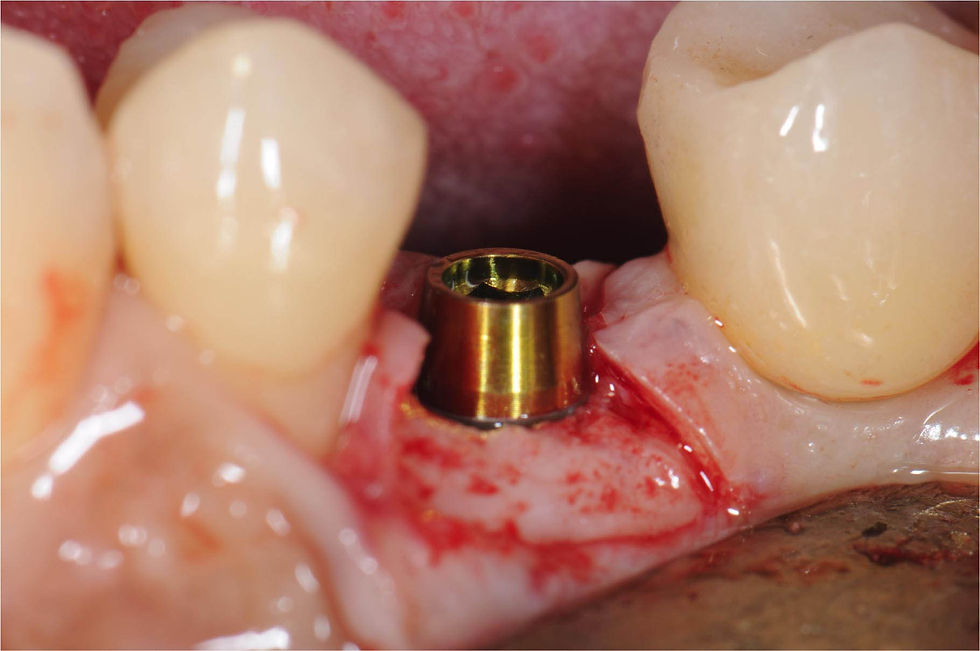
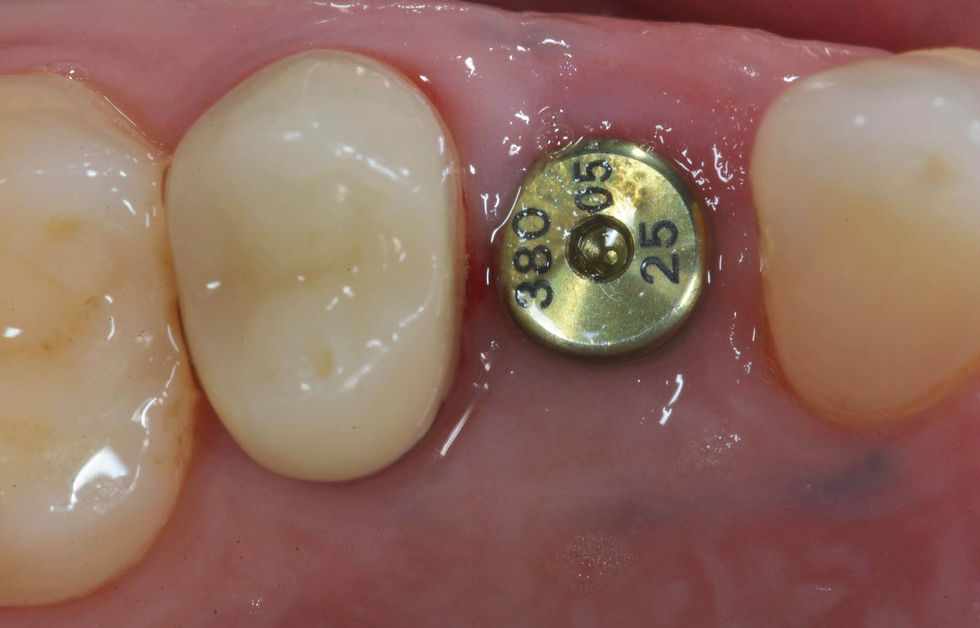
Gingival profile after removal of the Prama IN: the connection platform is at the level of the soft tissues, far from the bone for a length of 2.8 mm, which corresponds to the biological width.
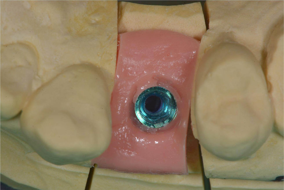
Laboratory phase with the analogue in the model.
Screw retained temporary crown in resin, made on Prama IN healing abutment, which maintains the same emergence profile and the same closure around the implant neck of the relative healing screw.
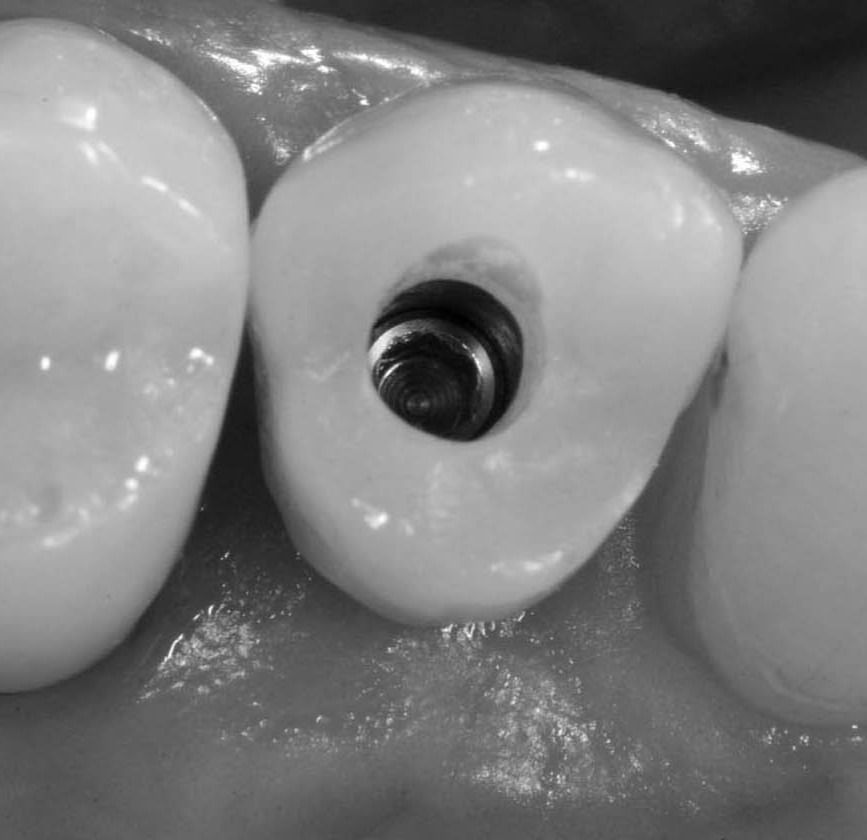
Temporary crown in place: occlusal view.

Temporary crown in place: clinical photo and radiograph.

Temporary crown in place: clinical photo and radiograph.
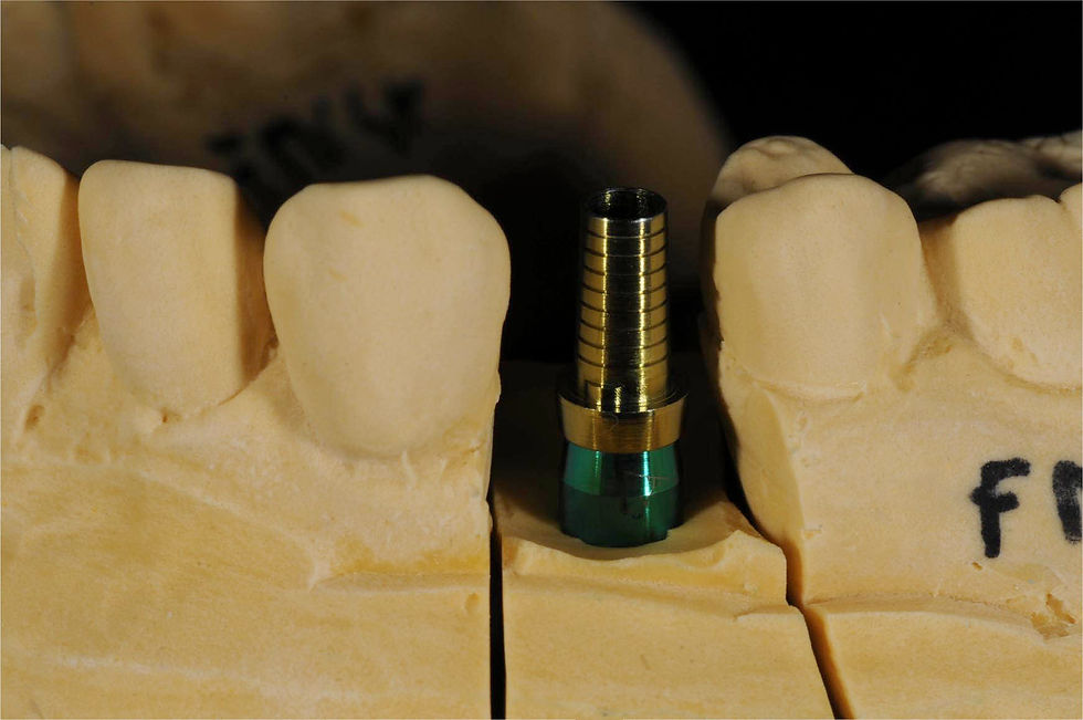
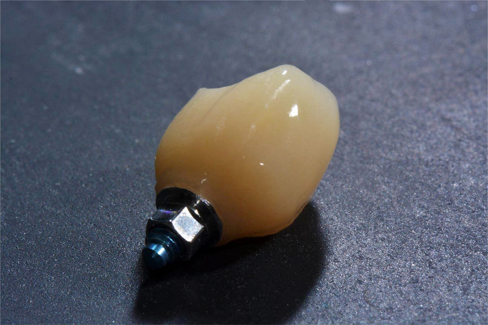
Laboratory phases to produce the definitive crown, made with Prama IN T-Connect.

Laboratory phases to produce the definitive crown, made with Prama IN T-Connect.

Adjustment and laboratory preparation of the T-Connect support.

Luting of the crown on the model.

Luting of the crown on the model.
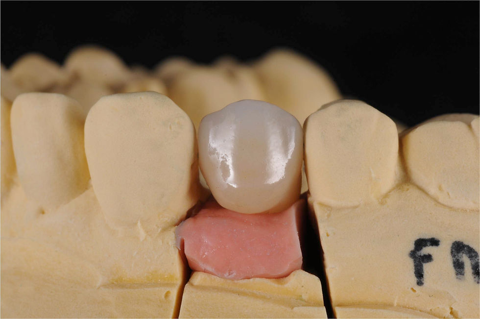
Finishing phases of the crown in the laboratory.

Finishing phases of the crown in the laboratory.

Radiographic follow up at 12 months.

Clinical follow up at 12 months.